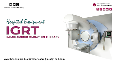
What is Image-guided radiation therapy?
Image-guided radiation therapy (IGRT) is the usage of imaging during radioactivity treatment to advance the exactness and correctness of therapy distribution. IGRT is consumed to treat cancers in parts of the form that interchange, such as the lungs. Radioactivity treatment machines made by IGRT Manufacturers are furnished with imaging know-how to permit your medic to copy the growth before and through therapy. By relating these imageries to the orientation imageries taken during replication, the patient's location and/or the radioactivity rays may be attuned to more exactly mark the radioactivity amount to the growth. To aid bring into line and mark the radioactivity gear, some IGRT actions may use fiducial indicators, ultrasound, MRI, x-ray imageries of the bone edifice, CT image, 3-D body exterior charting, electromagnetic transponders, or tinted ink summonses on the membrane.
If
you are to experience IGRT, your medic will be inclined to practice CT skimming
to steer a remedy imitation sitting and to generate orientation imageries.
Extra imaging procedures, such as MRI or PET image, maybe expended to aid
regulate the precise outline and site of your growth, and a singular expedient
may be shaped to aid you to uphold the same meticulous location during each
action. Your medic will give you exact directions founded on the kind of
examination being done.
In
IGRT gadgets that convey radioactivity that is provided by IGRT Suppliers, are
armed with singular imaging knowledge that permits the doctor to picture the
growth directly before or even through the time radioactivity is transported,
while the patient is located on the dealing table. Using dedicated mainframe
software, these imageries are related to the positioning imageries taken during
imitation. Any essential changes are completed to the patient's location and/or
radioactivity rays to more exactly mark radioactivity at the growth and evade
fit adjacent flesh.
In
IGRT, the gear used for taking images is affixed on or constructed into the
apparatus bought from IGRT Dealers that
transports radioactivity, such as a direct accelerator. Imaging gear may also
be fitted in the healing room Occasionally, IGRT is done by a sensor in the
room which paths gesture by confining indicators on the exterior of a patient,
or electromagnetic transponders positioned inside the patient.
Womenfolk
must always notify their doctor or engineer if there is any likelihood that
they are expectant or if they are breastfeeding their toddler. Patients with
pacesetters or slack metal in their forms should notify the healing team if MRI
is expended for imitation or IGRT.
For
particular IGRT actions, very minor indicators, which are called fiducial
indicators, or in some cases electromagnetic transponders may be located within
the form close or in the growth to aid the healing team to recognize the part.
They are typically positioned at least one week preceding the first
radioactivity healing cure. The patient's membrane also may be smeared or
tattooed with tinted ink to help bring into line and mark the radioactivity
gear. Patients with prostate melanoma who experience IGRT consuming ultrasound
must swig sufficient water around an hour before each therapy to keep their
bladder filled so that the prostate can be imaged or "understood" by
the ultrasound machine. At the start of each radioactivity treatment sitting,
the patient is prudently located directed by the spots on the skin outlining
the healing part. Expedients may be expended to aid the patient to uphold the
correct location. Imageries are then taken by consuming imaging apparatus made
by the IGRT Manufacturers that
is erected into the radioactivity distribution mechanism or fixed in the curing
room.
Some
IGRT methods necessitate patients to hold their inhalation for about 30 to 60
seconds. If IGRT necessitates fiducial indicators or electromagnetic
transponders within the form, these will be introduced into the form with a
pointer about a week preceding the imitation procedure.
On
all treatment days, contingent on the kind of IGRT being expended, doctors may
perform an x-ray, CT image, or ultrasound will be gotten preceding the healing.
The doctors or a radioactivity analyst will check the imageries and liken them
to the orientation imageries taken during replication to make location
alterations. The patient might be relocated and extra pictures may be taken.
After any vital modifications are finished to tie with the patient's location
introduction, radioactivity treatment is transported.


Comments
Post a Comment