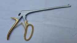
How are Casper Surgical Instruments useful in anterior cervical discectomy?
 |
| Casper Surgical Instrument |
A Casper Surgical Instrument permits surgeons to tug back, grip, and withdraw delicate tissues, such as the vertebral nerves and the spines during an anterior cervical discectomy. Anterior cervical discectomy and fusion (ACDF) is a kind of neck operation that comprises eliminating an injured disc to release spinal cord or nerve core pressure and ease conforming discomfort, faintness, numbness, and stinging. A discectomy is a system of medical decompression, so the process may also be named an anterior cervical decompression.
The
operation is advanced through the frontal, or obverse, of the cervical backbone
(neck). The disc is then detached from between two vertebral mandibles. A
synthesis operation is done at the same using equipment supplied by Casper
Surgical Instrument Suppliers time
as the discectomy process to steady the cervical section. A synthesis includes
enlisting bone shoot and/or grafts where the disc initially was in instruction
to deliver solidity and forte to the area. While this operation is most usually
completed to treat a characteristic cervical herniated disc, it may also
be done for cervical deteriorating disc illness. It is also usually done
to eliminate bone shoots (osteophytes) produced by arthritis and to ease the
indications related to cervical vertebral stenosis.
An
ACDF is completed with a frontal method, which means that the operation is
completed through the obverse of the neck as contrasting to over the posterior
of the neck. This method has several distinctive compensations:
- Straight admission
to the disc. The
forward approach permits the shortest conception of the cervical discs,
which are typically tangled in instigating the stenosis, vertebral cord or
nerve density, and indications. Elimination of the discs consequences in
shortest nerve and vertebral cord decompression. The forward method can
deliver admission to nearly the entire cervical backbone.
- A lesser amount of
postoperative discomfort. Vertebral column surgeons often favor
this method because it delivers access to the backbone through a
comparatively simple trail. The patient inclines to have less incisional
discomfort from this method than from a subsequent operation. After a skin
cut is completed in the obverse of the neck, only one reedy stunted muscle
needs to be bowdlerized, after which anatomic planes can be shadowed right
down to the backbone. The restricted volume of muscle separation or
partition helps to limit postoperative discomfort following the spine
operation.
While
an ACDF is the most usually achieved process for the management of cervical
disc pathology, a fresher process called a cervical synthetic disc standby, is
also obtainable. The general technique for an ACDF operation includes the
subsequent steps:
The
skin cut is one to two inches and is being made on the left or right-hand side
of the collar. The cut is typically made parallel within a usual skin crinkle,
though infrequently a more perpendicular cut is used for multilevel
circumstances. The reedy muscle under the skin is then divided in line with the
skin cut, and the level between the sternocleidomastoid muscle and the band
muscles then arrives. Subsequently, a level between the trachea/gullet and the
carotid cover arrives. A reedy fascia (flat coatings of rubbery flesh) shelters
the backbone (pre-vertebral fascia) and is then divided away from the disc
space.
Fluoroscopy
delivers an x-ray appearance of the back during operation and is used to settle
that the spine surgeon is at the precise disc level of the backbone. After the
precise disc space has been recognized, the disc is then detached by first
wounding the external annulus fibrosis (rubbery ring about the disc) and
eliminating the core purposes (the indulgent internal center of the disc). With
an anterior cervical discectomy, the complete disc is detached. The gristle
endplates on the vertebral frames are also detached to disclose the hard
cortical bone beneath.
Partition
is carried out from the obverse to the back of a tendon called the later
longitudinal tendon, which rests amid the disc and the vertebral cord. Often
this tendon is mildly detached to allow admission to the vertebral canal to
eliminate any disc solid that may have extruded through the tendon, which may
be backing to vertebral stenosis. The uncinated procedures—which are shares of
the lesser vertebral mandible on either side that help form the limits of the
disc—are characteristically at least moderately detached as well. This is
typically where osteophytes (bone branches) need to be detached to release
vertebral cord or nerve root firmness. The partition is often done using an
operating microscope or telescopic loupes to help with imagining the smaller
anatomic configurations.
Patients
characteristically go home the matching day as the anterior cervical discectomy
and fusion or after one night in the infirmary. Most patients recuperate within
about 4 to 6 weeks, and some can restore to most run-of-the-mill actions after
a few days or weeks. For the synthesis to be fully ripe— purporting that it
reconciles into one rock-hard, robust bone—may take a complete 12 to 18 months.
Patients should deliberate pertinent action limitations and reintegration with
their surgeon, as they will differ contingent on the person's complete health,
cervical back state, and the surgeon's skill. If you are looking for Casper
Surgical Instrument Dealers, please log into Ozahub.


Comments
Post a Comment